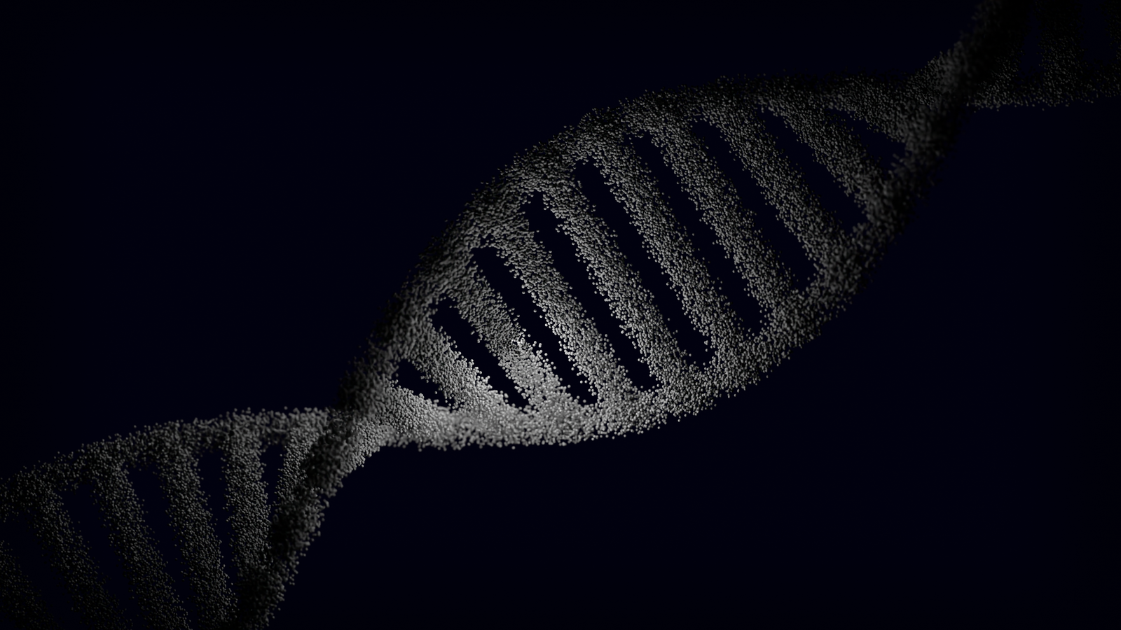
In May 1952, in a cellar laboratory at King’s College London, a graduate student named Raymond Gosling pointed a Philips micro-camera at a single fibre of hydrated DNA and let it soak up X-rays for roughly 62 hours. The plate he pulled out carried a sharp black cross — four bold smears in a diagonal X — and that pattern, catalogued as Photograph 51, was the fingerprint of a double helix. James Watson and Francis Crick were still shuffling cardboard cutouts in Cambridge when the picture that would decide the race was already sitting in a drawer at King’s.
The photograph was taken by Gosling under the direction of Rosalind Franklin, a 31-year-old crystallographer who had spent years in Paris perfecting the technique of firing X-rays through matter and reading the shadows. According to a detailed account in Genetic Engineering and Biotechnology News, the DNA fibres she was photographing had been supplied by the Swiss biochemist Rudolf Signer, drawn out into thin threads and coaxed into the hydrated “B” form by careful control of humidity in the sample chamber.
The cross on the plate was unmistakable. It also sat, for months, unused.
What a 62-hour exposure actually looked like
The camera itself was small enough to sit on a bench. Franklin had helped modify it, tilting the specimen, tightening the collimation, and rigging a system that bubbled hydrogen through salt solutions to hold the fibre at a specific humidity. The X-ray beam was a pencil of hard radiation aimed at a bundle of DNA barely thicker than a human hair. Almost all of the beam passed straight through. A tiny fraction bounced off the regular spacings of atoms inside the fibre and landed on a photographic plate behind it.
To collect enough of those scattered photons to build a readable image, the plate had to sit in front of the beam for days. Different sources give slightly different numbers — the play Photograph 51 and the King’s College archive have cited exposures of over 100 hours across a series of shots, while the single plate labelled Photo 51 is generally described as a roughly 62-hour capture. Either way, this was not a snapshot. It was a slow accumulation, closer in spirit to a long-exposure astronomical plate than to anything a modern camera does.
The exposure also came at a cost. Franklin worked without the shielding that would be standard in any X-ray lab today, and her ovarian cancer at age 37 has been widely attributed to that prolonged radiation exposure.
Reading a cross as a helix
A helix, spun in three dimensions and hit with a coherent X-ray beam, throws a very specific diffraction pattern: an X of bright spots, with the arms of the X made of layer lines whose spacing encodes the pitch of the spiral, and gaps at particular positions that encode the number of strands. Crystallographers had worked this out mathematically in the years before 1952. The theory was on paper. Nobody had ever seen it appear so cleanly in a biological molecule.
When Watson finally saw a print of Photo 51 in late January 1953, handed to him by Maurice Wilkins without Franklin’s knowledge, the pattern read like a sentence in a language he had just learned to parse. Watson later described his dramatic reaction upon first seeing Photo 51, writing that he was immediately struck by its significance, a line repeated in nearly every account of the affair, including the research briefing prepared for the West End play in which Nicole Kidman played Franklin.
The X told him it was a helix. The spacing between the layer lines told him the pitch was about 34 ångströms. The missing fourth layer line hinted that it was a double helix, two strands wrapped around each other and offset. Watson was not a trained crystallographer, but he had absorbed enough of the theory from Crick to know exactly what he was looking at.
The cardboard model in Cambridge
At the Cavendish Laboratory, Watson and Crick had spent the previous year effectively banned from model-building. Their first attempt in 1951, a triple helix with the phosphate backbones on the inside, had been dismantled during a day trip by Franklin, who pointed out that the water content was wrong by an order of magnitude. Their boss Sir Lawrence Bragg had told them to stop and let King’s handle DNA.
By early 1953, with Linus Pauling in California circulating a (wrong) triple-helix paper of his own, Bragg relented. Watson and Crick got their brass templates and cardboard cutouts back out. What they still lacked was hard experimental numbers: the pitch of the helix, the diameter, the density of the fibre, the way the strands were oriented relative to each other.
Two pieces of Franklin’s work supplied those numbers. The first was Photo 51 itself. The second was a Medical Research Council progress report that Max Perutz passed to Crick, which contained Franklin’s precise unit-cell measurements of the B form and, critically, her deduction that the two chains ran in opposite directions — antiparallel. The MRC report was not marked confidential, and nothing was technically stolen, but Franklin was never told her data had been shared.
Armed with her measurements, Watson found the base-pairing rule — adenine to thymine, cytosine to guanine — on a Saturday morning in February 1953, shuffling cardboard shapes on a desk in the Cavendish. He and Crick went to the Eagle pub for lunch. The model went up over the following days.
Why Franklin filed the photograph away
The strangest part of the story is that Franklin herself had seen Photo 51 months before Watson did and had chosen to set it aside. She was more interested in the crystalline A form of DNA, which produced denser, more information-rich patterns that she believed would eventually yield a full atomic structure by rigorous calculation rather than by guesswork with cardboard.
She distrusted model-building as a shortcut. The Cambridge duo’s 1951 flop had confirmed that view. Franklin wanted the answer to fall out of the mathematics, not out of a lucky arrangement of paper cutouts. It was a defensible scientific instinct that happened, in this case, to be the wrong one for winning a race.
By late 1952 she was also preparing to leave King’s for Birkbeck College, worn down by an environment where women were barred from the senior common room and she was treated as subordinate rather than an independent researcher. As she packed, Gosling gave the print of Photo 51 to Wilkins almost as a keepsake. Wilkins carried it to Cambridge.
Three papers in one issue of Nature
The published record kept the appearance of civility. In April 1953, Nature ran three back-to-back papers: Watson and Crick proposing the double helix, Wilkins and colleagues presenting supporting X-ray data, and Franklin and Gosling publishing Photograph 51 and its analysis under a technical scientific title.
Franklin’s paper appeared third and read like a confirmation of the Cambridge model, even though her data had been the model’s foundation. She added a final sentence noting that her ideas aligned with Watson and Crick’s proposal. Watson and Crick’s paper thanked her with a footnote vaguely acknowledging her unpublished results. The wording understated the debt to the point of misdirection.
In 1962, the Nobel Prize in Physiology or Medicine went to Watson, Crick and Wilkins. Franklin had died of ovarian cancer four years earlier at 37, and the Nobel is not awarded posthumously. Her name was not on the citation. Recent obituaries revisiting the discovery, including Ars Technica’s account of Watson’s death in 2025 at age 97, describe the omission as a permanent asterisk on the story.
The long correction
Watson’s 1968 memoir The Double Helix made things worse before they got better. He referred to Franklin as “Rosy,” a nickname she disliked, portrayed her as prickly and hyperemotional, and cast her as an obstacle rather than a source. The book was a bestseller and shaped a generation’s understanding of the discovery.
The rehabilitation started with Anne Sayre’s 1975 biography and continued with Brenda Maddox’s Rosalind Franklin: The Dark Lady of DNA in 2002. Maddox’s husband, the former Nature editor Sir John Maddox, later said that had he been editing Nature in 1953, he would have questioned the vague acknowledgement of Franklin’s data and might have insisted she be listed as a co-author. That quote appears in the GEN retrospective on Franklin’s centenary.
By the time Nature ran a follow-up assessment of the affair, historians had largely settled on a firmer verdict: without Photograph 51 and the MRC report, Watson and Crick would not have built the model when they did, and possibly not at all before Pauling corrected his own errors. Professor Lynne Osman Elkin has argued that Watson and Crick could not have determined the structure before Franklin’s draft was published without access to her data.
A pattern in twentieth-century labs
Photograph 51 belongs to a small class of accidents and instruments that produced the twentieth century’s biggest biological jumps. It was a physical object — a piece of glass with silver grains rearranged by X-rays — that carried more information than anyone in the room could quickly extract, and it changed hands in ways nobody had properly documented.
There is a family resemblance to other moments Space Travel has covered. When Alexander Fleming returned from holiday in 1928 to find a mould-cleared ring on a Staphylococcus plate, the discovery lived first as an image before it lived as a theory. When John Snow mapped cholera deaths around a Soho pump in 1854, the argument was carried by dots on a street plan. Photo 51 fits the same category: a single piece of visual evidence that made an abstract claim suddenly undeniable to anyone trained to read it.
The difference in Franklin’s case is that the person who produced the evidence never got to present it as her own before somebody else had built a model around it. The photograph moved faster than the paperwork.
Where the plate is now
Photograph 51 itself survives. The original plate and the Philips micro-camera used to make it are held in the King’s College London archives, and researchers preparing the recent opera about Franklin’s life have been shown both. Kidman was allowed to handle the camera during her research for the 2015 play. Brian Sutton, professor of molecular biophysics at King’s, has described how visitors reach for the equipment first and the photograph second, as if the object that made the image somehow matters more than the image.
The European Space Agency named its ExoMars rover Rosalind Franklin in 2019. Its mission is to drill into Martian regolith looking for organic molecules — the same class of long-chain carbon structures whose most famous member she had photographed 70 years earlier in a King’s basement. The rover carries her name across roughly 300 million kilometres of interplanetary space, further than any tribute etched on a building in Bloomsbury.
The plate itself, meanwhile, sits in a climate-controlled archive not far from where it was exposed. A person allowed to lift it out of its folder sees, through the yellowing emulsion, four dark smudges arranged in an X. It took 62 hours of X-rays and one steady graduate student to put them there. It has taken most of a century to attach the right name to what they showed.